Home › General Account of Butterflies › II. the Structure of Butterflies: Lepidoptera
II. the Structure of Butterflies: Lepidoptera
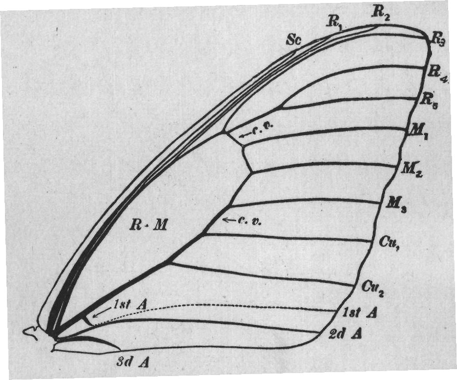
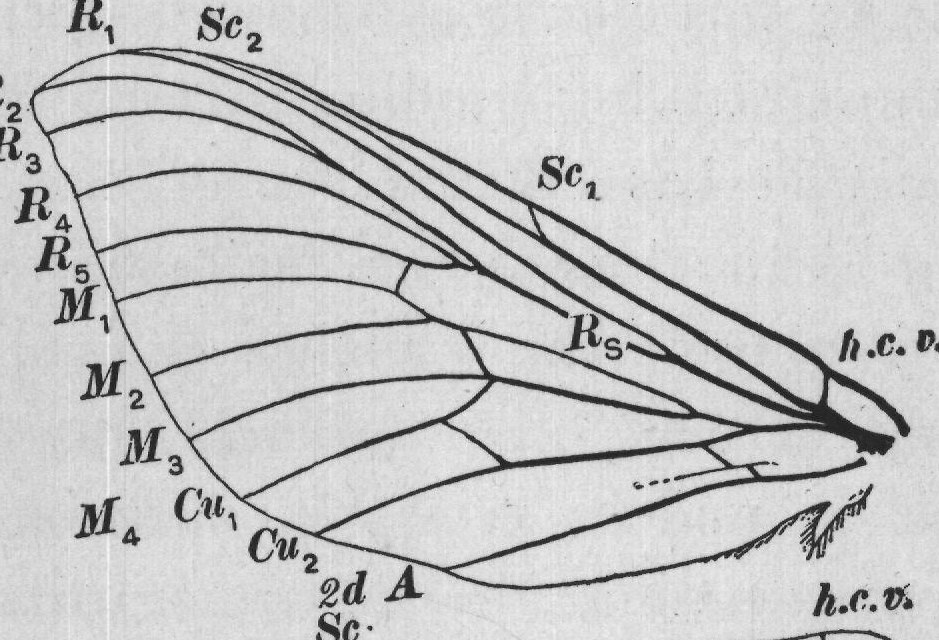
It is only in a very few Lepidoptera that there is any indication of the existence of vein M₁₄. In nearly all moths and in all butterflies media appears to be only three-branched; and it is customary to regard the vein labeled M₁₄ + Cu, in the above figure as merely cubitus-one. For the sake of simplicity this course is followed in this work. 4 I The most striking modification of the primitive type that has taken place in the wings of butterflies is the loss of the main stem of media and the joining of the branches of media to the veins on either side. Thus vein M, appears to be a branch of radius; vein M₃, of cubitus; and vein M, is sometimes joined to radius and sometimes to cubitus. That this change has taken place is easily seen by comparing the venation of Sthenopis (Fig. 6) with that of the various butterflies figured here. 2 Not only has the main stem of media been lost, but in nearly all wings of butterflies one or two of the three anal veins have disappeared. A careful study has shown that in the reduction of anal veins in the Lepidoptera the first anal vein is the first to disappear, and the third anal vein is the next to go.
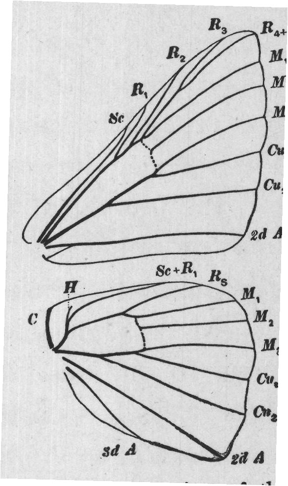
The number of the branches of a branched vein is often reduced by the growing together, or I2 coalescence, of adjacent veins. Thus in many butterflies radius of the fore wings instead of being five-branched is only four-branched or even threebranched (Fig. 7). In a case of this kind the designation R4+5 indicates that veins R, and R , have grown together to form a single vein.
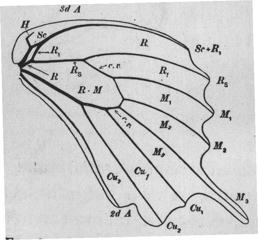
In the hind wings of all butterflies the radius differs greatly from that of the fore wings. By referring to the venation of Sthenopis (Fig. 6) it can be seen that at the first forking of radius the vein is divided into two unequal parts; one of these is vein R₁, the other gives rise to the remaining four branches of radius. This S C H Se R sd A 4 R3 R4+5 R2 M M2 M Cu, Cu 2d A Sc+R, Rs M. 1 M 2 M Cu 20 zd A FIG. 7.-Venation of the wings of a metal-mark. second part is termed the radial sector, and is labeled R, in the figures. In all butterflies the branches of the radial sector of the hind wings all coalesce so as to form a single vein, and vein R, coalesces with the subcosta. This is well shown in shown in the hind wing of Papilio (Fig. 8). It will be observed that in this wing vein R, soon after its separation from vein R, joins vein Sc and the S two extend to the margin of the wing as a single vein, which for this reason is marked Sc+R,. It will also be seen that the radial sector extends to the margin of the wing as an unbranched vein, R-M 1st A 3d A H Sc R R R S Sc R R2 R₃ R R ٢..
M M2 M C. D.
Cu, Cu2 1st A 2d A Sc+R, Rs R R RM M M M M M, Cu, 2d A Cu 1 Cu2 Cu 2 4 M 3 FIG. 8.- Venation of "the wings of a of markings, it is swallow-tail. marked R..
In some butterflies a short spur extends from the subcosta near the humeral angle of the wing; such a spur is termed the humeral (Fig. 7, H). vein The thin spaces of the wings which are bounded by the veins are called cells. In descriptions of wings, especially in indicating the location of markings, it is often desirable to refer to one or more cells. It is necessary, therefore, to have a nomenclature of the cells of the wing, as well as of the wing-veins.
Having named the wing-veins, the simplest possible method of designating the cells of the wing is to apply to each the abbreviation of the name of the vein that forms its front margin.
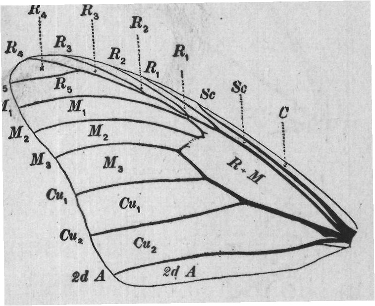
In Figure 9 the veins are designated by letters at the margin of the figure; the cells by letters within the figure or at the ends of the dotted lines.
Near the center of the basal half of the wing there is a large cell RA R3 which is bounded in R2 R R3 front by the main R2 R R R 5 R5 Sc Sc stem of radius and M M C 1 M M2 2 which for this reason M M3 3 R+M might be called cell Cu R. But this cell is Cu2 2 Cu, Cu2 really composed of 2d A 2d A two cells, which have FIG. 9.-Fore wing of a butterfly with been thrown together the veins and cells named. by the fading out of the main stem of media.
For this reason this cell is designated as cell R+M. This is the discal cell of most writers on the Lepidoptera.
The details of the venation of the wings can be seen best, in Lepidoptera, on the lower surface of the wings; as on this surface the veins are not so obscured by scales as on the upper surface. If necessary the scales can be removed from a small part of a wing with a small brush in order to expose the veins. A drop of chloroform applied to a wing causes the veins to be more prominent for a moment; the chloroform soon evaporates and leaves the wing uninjured.
At the base of each fore wing there is a prominent scalelike appendage; these are known as the patagia.
The third and last region of the body, the abdomen, requires little discussion in this place. It consists of several ringlike segments, and bears appendages at the caudal end connected with the organs of reproduction. The forms of these appendages vary greatly in the different species, consequently much attention is devoted to them in the more technical works.