Home › General Account of Butterflies › II. the Structure of Butterflies
II. the Structure of Butterflies
The body of a butterfly consists of three regions, which are known as the head, the thorax, and the abdomen. The head is the first of the three regions; the thorax, the intermediate; and the abdomen, the last.
The head bears the eyes, the antennæ, and the mouth-parts.
The eyes are two in number, one on each side of the head. They are easily recognized by their position and hemispherical form. But when they are examined with a lens they present a very different appearance than do the eyes of man; each eye being composed of a large number of little eyes, or ommatidia as they are termed. As the ommatidia are closely massed together, the outer surfaces of each is hexagonal in outline like the cells of honeycomb (Fig. 3).
Eyes of this type are termed compound.
Many insects have simple eyes, or ocelli, in addition to compound eyes; but ocelli are very rarely found in butterflies.
The antenna are the long, more or less threadlike appendages that project from the upper part of the head; they are what children are apt to call the horns of the butterfly.
Each antenna consists of many segments or ringlike divisions.
The antennæ are supposed to bear the organs of smell. In pound eye, greatly butterflies the terminal segments of the antennæ are en- FIG. 3.-Part of a commagnified. larged so as to form a club.
The mouth-parts of butterflies consist chiefly of a pair of palpi and the sucking organs, maxilla. The palpi are the jointed organs that project forward from the lower side of the head. They vary greatly in length in different families, and vary in the relative length of their segments, so that use is made of them in the classification of butterflies. The maxillæ are greatly modified jaws, which are so lengthened that they have lost all resemblance to the jaws of biting insects.
Each maxilla is furnished with a groove, and the two maxillæ are so fastened together that the two grooves form a tube through which liquid food is sucked. When not in use, the maxillæ are coiled between the palpi.
The thorax bears the organs of locomotion, the legs and the wings.
The legs are six in number. Each leg consists of a series of segments. The basal segment, that by which the leg is attached to the body, is the coxa; next is a small segment, the trochanter; then follows the principal segment of the leg, the femur; the next approaches the femur in size, and is the tibia; the remaining segments constitute the foot or tarsus. The last segment of the tarsus usually bears a pair of claws.
The wings are four in number and are always present in adult butterflies. In many species of moths the wings are wanting in one sex; but this is true of no butterfly.
In the study of the classification of butterflies much use is made of the variations in the structure of the wings. This is also true in the study of any of the groups of winged insects; but in the Lepidoptera, where the body is covered with a dense clothing of scales which hides from view most of the distinguishing characteristics used in the classification of beetles and other comparatively naked insects, the structure of the wings presents an even larger proportion of the easily available criteria for separating the order into its subdivisions .
It is essential, therefore, that the student of butterflies should learn at the outset the more important facts regarding the structure of the wings, and become familiar with the terms that have been applied to the different parts of a wing.
Fortunately it is an easy matter to do this.
The two pairs of wings are designated as the fore wings and the hind wings respectively.
Some writers on butterflies term the fore wings the primaries, and the hind wings the secondaries.
The wings are more or less triangular in outline; a wing, therefore, presents three margins : the costal margin, or costa (Fig. 4, a−b); the outer margin (Fig. 4, b−c); and the inner margin (Fig. 4, c-d).
Fig . 4, c-d).
The angles limiting these margins have also received names. The angle at the base of the costal margin (Fig. 4, a) is the humeral angle; that between the costal margin and the outer margin (Fig. 4, b) is the apex of the wing; and the angle between the outer margin and the inner margin (Fig. 4, c) is the anal angle.
The wings are large membranous appendages, which are thickened along certain lines. These thickened lines are termed the veins of the wing; and their arrangement is described as the venation of the wings.
A study of the wings of all orders of winged insects has shown that there is a striking uniformity in the more general features of the venation of the wings of the more generalized or "lower" members of the different orders; while in the more specialized or "higher" a a d b C d C b FIG. 4.-Margins and angles of a wing. members of each order this generalized type of venation is more or less modified.
An investigation of the various ways in which this generalized type of wing venation has been modified and of the varying degrees of these modifications has contributed much to our knowledge of the relationships of the different groups of insects.
These studies have shown that all winged in sects have doubtless descended from a common winged ancestor. And although we do not know the exact form of this primitive winged insect, which lived during the Silurian age, we may infer that those structural features that are common to the generalized members of the different orders of winged insects have been inherited from this common ancestor.
The features of the wing-venation which are commonly present with the generalized members of the different orders of winged insects, and which we therefore infer were possessed by the primitive winged insect, are represented in Figure 5.
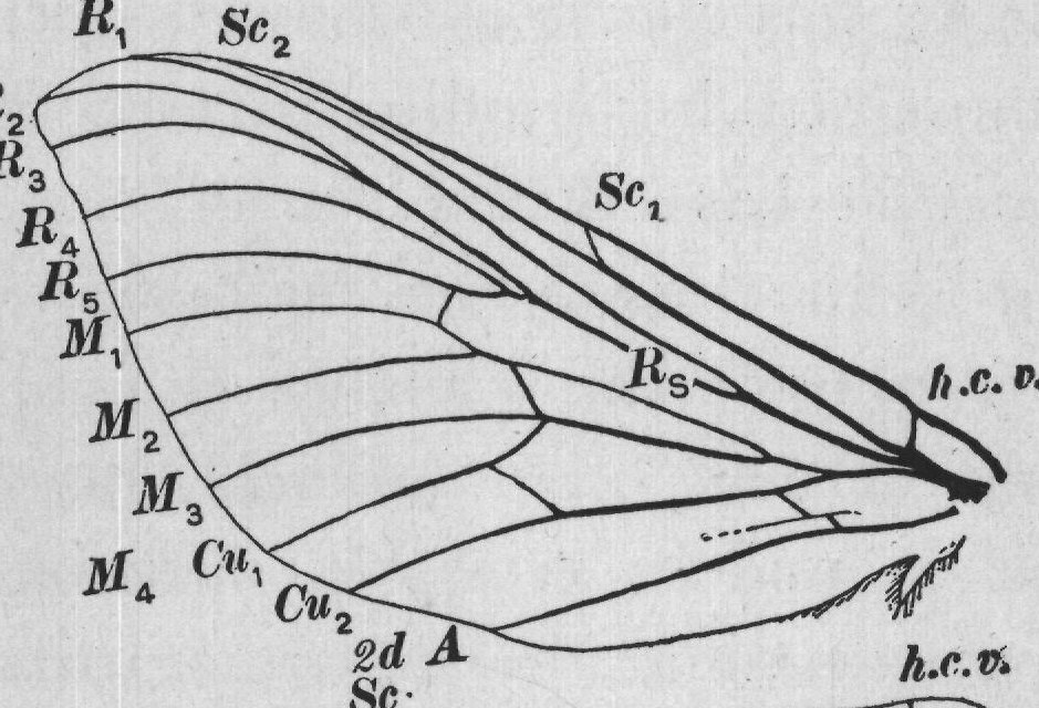
From this hypothetical primitive type of wing-venation there can be derived, by methods of modification of which we have many illustrations among living insects, all the forms of venation of insect wings known. The venation of the wings of butterflies is one of the more specialized types of wing-venation, and one which can not be understood by the study of the wings of butterflies alone. It is necessary, therefore, to lead up to the explanation of this type by describing more simple or less modified types. We will describe first the hypothetical primitive type and then point out the ways in which this type has been modified in the Lepidoptera. Taking the hypothetical type (Fig. 5) as an illustration, we see that the veins of the wings can be grouped under two heads: first, longitudinal veins, those that normally extend lengthwise of the wing; and second, cross-veins, those that Sc, Sc₂ R, R2R?
C h -C- SCR M Cu r-m -m--cu m R₄ 4 R5 1 AMM 2 2d A 3d A 1st A Cuz M Cu M a FIG. 5.-Hypothetical venation of the primitive winged insect. extend transversely from one longitudinal vein to another.
The names that have been applied to the longitudinal veins, beginning with the one nearest the costal margin of the wing, are costa, subcosta, radius, media, cubitus, first anal, second anal, and third anal. In descriptions these veins are often designated, as they are in Figure 5, by the following abbreviations of these names: C, Sc, R, M, Cu, Ist A, 2d A, and 3d A.
Beginning with subcosta, the four veins that traverse the middle portion of the wing are branched; the subcosta divides into two branches, the radius into five, the media into four, and the cubitus into two. In this primitive type the costa and the three anal veins are not branched.
In designating the branches of a forked vein they are numbered, beginning with the one nearest the costal margin of the wing. Thus, the first branch of radius is designated as radius-one; and for this term the abbreviation R₁ is used.
In some insects there are very many crossveins, but it is believed that the greater number of these have been developed secondarily. There are, however, a few cross-veins that are so con. stantly present among generalized insects that we feel warranted in believing that they were present in the wings of the primitive winged insect. These are represented in Figure 5, and are desig. nated as the humeral cross-vein (Fig. 5, h); the radio-medial cross-vein (Fig. 5, r-m); the medial cross-vein (Fig. 5, m); and the medio-cubital crossvein (Fig. 5, m-cu). In Figure 6 is represented the venation of the wings of Sthenopis, a moth, which is one of the most generalized of the living Lepidoptera. Here is found quite a close agreement in venation with IO that of the hypothetical type. The more important modifications are the following: The costa forms the costal border of the wing, and does not appear as a distinct vein. This is the case with nearly all insects; but in many pupæ the costa is distinct, and it is only in the later R 2 R3 R 4 R R 5 M1 1 M2 2 M 3 SC2 Ma Cu 4 R2 R 3 1 Cu2 2d A R Sc Sc, Rs RS 1st A 2 h.c. D. h.c.v. 3d A 2d A FIG. 6.-Venation of the wings of Sthenopis. stages of the development of the wings that it coincides with the costal margin. 4 In the hind wings, veins M, and Cu, unite for a short distance, and then separate; in the fore wing these veins unite and remain united throughout the remainder of their length (Fig. 6, M₄ + Cu,).