Home › Ancient Salamanders › Ancient Salamanders: Mastodonsaurus
Ancient Salamanders: Mastodonsaurus
Turning to their skulls, there is one feature that is quite peculiar to the group and rarely seen in reptiles, living or extinct, viz. that the whole of the upper surface of the skull behind the eye-holes is covered in by a complete roof of bone. Hence some authorities call them Stegocephali,¹ or "roof-skulled." This is clearly shown in Fig. 16, representing the huge skull of Mastodonsaurus. Some fishes have skulls thus roofed over. But the skull of a lizard or a crocodile is very different, showing a long open channel behind each orbit, and another one lower down at the side of the head. Another important character in the labyrinthodont skull is the presence of two condyles, instead of only one, as in all true reptiles and birds. The word condyle 2 signifies a knuckle, and is applied chiefly to the surface or surfaces by which the skull joins on to, or articulates with, the first vertebra of the neck. All the mammalia have skulls articulating by two condyles-a fact which would seem to imply that mammals branch off from the amphibia, and not from reptiles. Again, the bones of which the skull is composed are nearly always covered with a network of grooves, or "canals," doubtless intended for the attachment of hard, horny, and bony scales or scutes.
Greek-stegos, roof; cephalos, head. 2 Greek-condulos.
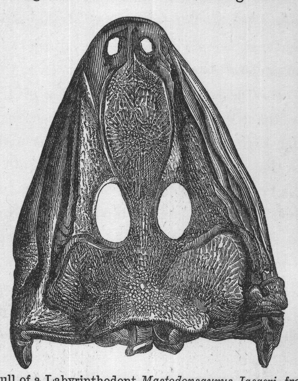
Before we leave the skull there is one other very interesting point that should not be omitted. If the reader will refer to the skull of Mastodonsaurus (Fig. 16), he will see, just on the middle line and near the base, a small round hole. It lies over the brain-cavity, and most probably represents the site of a small third eye. Whether this little extra eye was capable of receiving impressions of light, and so of communicating them to the brain FIG. 16.-Skull of a Labyrinthodont Mastodonsaurus Jaegeri, from New Red Sandstone, Germany. as an ordinary eye does, is a matter of doubt; but in a certain very primitive little lizard now living in New Zealand, and known as the Tuatara (Sphenodon), there is a similar aperture overlying the rudiment of an eye now quite useless and lying down in the brain. This little eye in the Tuatara must at one time have been made use of, and the question arises whether the third eye of the Labyrinthodonts was similarly useless; but the chances are rather against the notion. All the lizards have this pineal eye. It is probable that we have here a relic from some much older and now lost form of lifepossibly a fish, but possibly also a still lower type. These rudimentary structures, of which Darwin speaks in his great work, The Origin of Species, always possess a high degree of interest for the palæontologist, as being survivals from very early days in the world's history. To the latter they are as interesting in their way as are ancient customs and old superstitions to a student of folk-lore or of ancient history.
We must also notice the forward position of the creature's nostrils as indicated by the two small openings at the apex of the skull (see Fig. 16). If we were to examine the under side of a skull such as that of the Mastodonsaurus, we should see that the bony palate is formed chiefly of two broad and flat bones, called the "vomerine," which generally support teeth. Now, this is a character not exhibited by the skull of any true reptile, and we must go to modern amphibia, such as our frogs and toads, to find a parallel. There is also a corresponding series of small teeth on the mandible of the lower jaw (see Fig. 25). The eyes often show the peculiar sclerotic plates seen so well in the "fish-lizards."
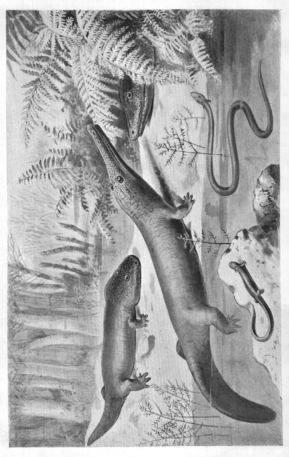
In studying an extinct vertebrate, or backboned animal, one of the first points to be considered is the nature of the vertebræ, or joints of the backbone. Now, in all fishes, and also in their distant cousin the Ichthyosaurus (fish-lizard), these bones have their articulating surfaces (centra) hollow, hence the popular term cup bones" applied to the vertebræ of the latter. Need we be surprised, then, to learn that the Labyrinthodonts possessed similarly cup-shaped vertebræ? This is one more link to connect them with fishes. In some of the more primitive and ancient 66 forms, the body of the vertebra was composed of three separate pieces, an arrangement peculiar to certain primitive fishes. Ever since an unfortunate restoration of a Labyrinthodont by Mr. W. Hawkins, it has been the custom-in popular works at least to represent the creature with a frog-like aspect. This restoration was based on the imperfect material of a good many years ago, and is therefore out of date. But later discoveries have shown that, instead of being frog-like, most of the Labyrinthodonts, if we could have seen them in the flesh, would have reminded us more of our modern little newts, or of the salamanders of hot countries. The tail was generally well developed, and the limbs were adapted quite as much for walking as for swimming.
Their chests were defended by three bony plates (see Fig. 17), which had grooves similar to those on the skull. But besides this, all the under side of the body was protected by a large number of scales, running in two directions, as shown in Fig. 17. In one or two instances the whole body was covered with a coat of mail, consisting of small bony scales. An example of this kind of skin is the Seeleya, from the Permian rocks of Bohemia, restored by Fritsch, and named by him after the late Professor H. G. Seeley, F.R.S. In size the Labyrinthodonts varied greatly, some being only an inch or two in length, others seven or eight feet, and perhaps more. Most of them had limbs, but in one or two cases these had apparently been dispensed with. It might naturally be asked, Did they undergo a series of changes similar to the metamorphoses of a common frog? One could hardly, perhaps, have expected an answer to this question; but, strange to say, some of the smaller forms from the petroleum shales (Gaskohle) of Bohemia, so carefully studied and described 1 Smit AMPHIBIANS OF THE COAL-FOREST AND PERMIAN PERIODS.
Actinodon.
Loxomma (Head of).
PLATE VII.
Ceraterpeton.
Dolichosoma.